
mon-fri 9-13 / 14.30-18.30
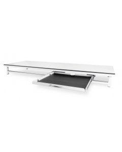
Veterinary radiographic table in steel (160x65 cm) with radiolucent HPL top. Silent and dry-running mobile drawer, 150 kg capacity.
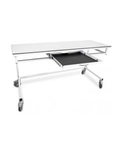
XR-SLIDE 2 steel veterinary radiographic table. X-ray transparent HPL top (160x65 cm). Dry drawer, 150 kg load capacity.
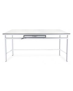
Veterinary radiographic table with radiolucent HPL top, mobile drawer for RX detector, steel structure and adjustable feet. Capacity: 150 kg.
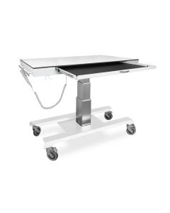
Veterinary radiographic table in steel with radiolucent HPL top. Adjustable height, detector drawer and 200 kg load capacity.
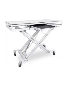
Veterinary radiographic table in steel with X-ray transparent top in HPL/polycarbonate. Battery-powered electric adjustment, load capacity 200 kg.
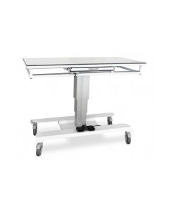
Steel veterinary x-ray table with radiolucent HPL top (135/160x65 cm). Lifting column and dry-running drawer, 200 kg capacity.
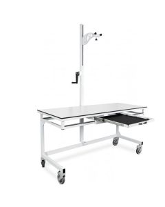
Veterinary radiographic table with radiotransparent HPL top, sliding drawer, adjustable support for portable RX units and braked wheels.
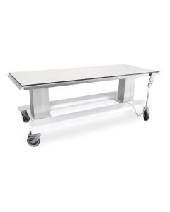
Veterinary X-ray table in steel with radiolucent HPL top (160x65 cm). Double electric column for height and tilt, 200 kg capacity.
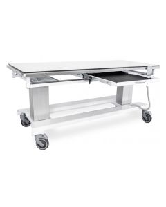
2-column fire-retardant veterinary radiographic table. Tilting top in radiolucent HPL (160x65 cm). Dry-running drawer, 200kg capacity.
Veterinary radiographic tables represent the central element in the setup of any clinical or hospital radiology room intended for veterinary medicine for small and medium animals. The choice of an appropriate diagnostic support not only affects the overall quality of the final radiographic image, but also directly influences the operator’s work ergonomics, safety against exposure to ionizing radiation, and patient restraint management. Within modern facilities, a well-structured radiological table must guarantee perfect integration with direct digital acquisition technology (DR) or indirect (CR), while offering mechanical robustness suitable for withstanding continuous daily stress and substantial body weights.
Technological evolution has transformed these elements from simple fixed support surfaces into true multifunctional dynamic systems, equipped with electro-mechanical movements, high-efficiency radiotransparent tops, and advanced centering systems. Correctly evaluating the load-bearing structure, surface coating materials, and receptor drawer locking mechanisms allows for optimizing examination execution times, reducing the need for repeated exposure and minimizing animal stress. The shape of the top and the presence of accessories dedicated to anatomical positioning are key factors for obtaining precise and diagnostic orthogonal projections, protecting the health of the spine and joints of the healthcare personnel involved.
The integration of a professional radiological system specifically for the veterinary field responds to the primary need to guarantee high diagnostic performance and maximum operational safety. Unlike human medical diagnostics, the veterinary patient does not actively collaborate in positioning, requiring physical or pharmacological restraint and extremely short dwell times. Radiological tables for veterinary use integrate mechanical solutions specifically designed to facilitate the loading of animals of different sizes, breaking down physical barriers thanks to variable height systems that minimize the lifting effort by healthcare operators and veterinary doctors.
In addition to the ergonomic aspect, the presence of a high radiotransparency work surface ensures minimum attenuation of the X-ray beam generated by the X-ray tube, preventing the formation of artifacts on images and reducing the overall dose required for correct exposure. The use of advanced materials such as high-density polycarbonate or radiotransparent structural laminates allows for maintaining excellent flexural rigidity without compromising the homogeneity of radiation transmission. This translates into superior image clarity, fundamental for identifying micro-fractures, mild pulmonary pathologies, or early-stage abdominal anomalies, elevating the general level of clinical performance provided by the facility.
The range of radiographic tables intended for veterinary diagnostics is divided into different construction configurations, designed to meet the various dimensional, operational, and economic needs of healthcare facilities. The fixed top variants with constant height constitute the ideal solution for radiology rooms with standard operations or limited spaces, where mechanical simplicity is combined with high stability and maintenance requirements reduced to a minimum. Such models are frequently equipped with sliding cassette or DR panel drawers along the longitudinal axis, equipped with integrated or interchangeable anti-scatter grids to optimize yield on thicker anatomical districts.
For high-workflow clinics and veterinary hospitals, tables with a 4-way floating top and electric or hydraulic elevation represent the reference performance standard. The movement of the top in the four spatial directions allows for centering the anatomical area of interest under the radiation beam without having to manually move the animal, a particularly delicate and painful operation in cases of severe trauma. The presence of motorized height adjustment facilitates autonomous access for large patients and prevents incorrect posture for the operator during positioning and physical restraint phases.
Selecting the ideal veterinary radiographic table requires careful prior analysis of clinical workflows, the prevailing type of patients treated, and the emission and detection technology already present in the facility. The first decisive factor lies in the dimensional and mechanical compatibility with the digital detector in use, whether it is a corded or wireless FPD (Flat Panel Detector), ensuring that the drawer below allows for secure housing of the device and perfect centering of the power grid. It is essential to evaluate the maximum useful capacity of the load-bearing structure, ensuring a load capacity suitable to safely support even giant breed dogs without flexion or vibration of the top.
Another critical aspect concerns the choice of surface materials and the methods for unlocking movements. The presence of foot controls for unlocking the floating top brakes allows the doctor or technician to keep both hands on the patient during the centering phase, increasing time efficiency before the shot. The resistance of surfaces to biological liquids and hospital-grade disinfectant detergents represents a fundamental requirement for preventing early corrosion of metal parts and degradation of seals, safeguarding the value of the investment in the long term.
All radiographic tables intended for use in the veterinary field must meet strict construction requirements regarding electrical safety, mechanical stability, and radiation protection, in compliance with current European directives and national regulations. Compliance with electromagnetic compatibility (EMC) requirements and the machinery directive guarantees the absence of interference with other electromedical devices present in the diagnostic room, as well as protecting the safety of the operator during the activation of lifting motors or electromagnetic locking systems.
From a radiation protection perspective, the structure of the table and the receptor drawer must provide adequate rear shielding in lead or equivalent material, aimed at limiting the scattered radiation below and fulfilling the obligations set by the radiation protection expert for the installation room. The adoption of certified components and the periodic execution of quality controls on mechanical performance and beam centering are indispensable prerequisites for maintaining the operational suitability of the system and avoiding sanctions during verification by the relevant supervisory bodies.
Explore all the products in the category and compare the available solutions to find the one best suited to your operational context.
The fixed table requires direct manual positioning of the patient under the x-ray beam. The model with a floating top, instead, allows the work surface to be gently translated in four directions, keeping the animal still and facilitating quick and accurate anatomical centering.
Tops made of high-density polycarbonate or high-pressure structural laminate guarantee a very low X-ray attenuation coefficient. This allows for reducing the delivered dose while maintaining high contrast quality of the diagnostic image obtained.
Modern tables have universal drawers with adjustable stops, designed to accommodate both corded and wireless standard-sized DR digital panels. The structure often includes a slot for inserting a removable anti-scatter grid.
Motorized height adjustment is strongly recommended in facilities that manage large animals. It allows the patient to step on autonomously when the top is lowered and offers the operator the possibility of working at an ergonomically correct height.
The table surfaces must be cleaned after each examination using non-corrosive hospital-grade disinfectants. Periodic maintenance includes checking electromagnetic locking systems, lifting cables, and the smooth sliding of the receptor drawer.
The lower part of the cassette drawer usually integrates a lead shielding sheet to absorb residual radiation. The overall compliance of the room must still be evaluated by the appointed radiation protection expert during the first installation.
Each model has a certified maximum capacity specified by the manufacturer, usually between 100 kg and 150 kg. It is essential to check the nominal load capacity to ensure structural safety during diagnostic procedures on mastiffs or giant breed dogs.
 English
 Italian
 French
 German
 Spanish